Vitreous humor is a transparent gelatinous mass between the lens and the retina that fills 80% of the eyeball space. It acts to transmit the light rays from outside to the retina, providing nutrients for the lens and retina. It also creates pressure to fix the lens shape, helping the lens to focus a sharp image on the retina. The vitreous humor is formed since one’s birth, stays forever, and should not be replaced, except by human action (Vitrectomy). Vitrectomy is a procedure to remove part of or entire vitreous humor to create better access to the retina allowing for the treatment of retinal diseases.
For those with retinal diseases, if their surgery has not been performed immediately, they should limit activities such as neither take a flight nor do vigorous activities. Besides their basic needs, they should lie down in bed, avoid too many activities.
- Have breakfast normally on the day of surgery (except for those with diabetes).
- Should not use alcohol or stimulants before the surgery.
- Continue using daily prescription drugs if any.
- Should not make up and use cosmetics, keep eyes and face clean on the day of surgery.
- Should not wear tight pullovers or shirts made of fur.
- Should not wear items such as jewelry, watches or hairpins into the operating room.
- Get enough sleep and stay comfortable before the day of surgery.
- Should not drive on the day of surgery.
Around 1 hour before the surgery, you shall be provided with
Around 1 hour before the surgery, you shall be provided with antibiotics, anti-inflammatory and pupil dilation every 3-5 minutes.
Step 1: Local anesthetic injection near the eyeball
The eye is cleaned with an antiseptic solution; then local anesthetic drops are given first to numb the surface of the eye and anesthetic injection is given near to the eye, but not the eyeball itself. You may first feel stinging or pressure which usually lasts within 3-5 minutes.
Step 2: Perform cataract surgery
In some cases, you need to get a cataract surgery together with your vitrectomy.
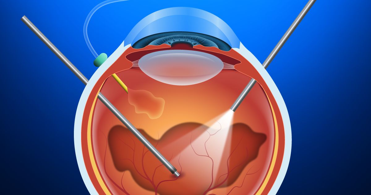
Step 3: Create an access to the vitreous body
Trocar – an instrument to create cannulas to the vitreous body through the cornea. An infusion line is created at one cannula to maintain the eyeball shape, a light source will be placed at another cannula, a vitreous cutter or a laser tip will be placed at the other (if necessary).
Step 4: Cut and remove the vitreous humor
First, the surgeon will cut and remove the vitreous humor. And then, additional procedures shall be carried out depending on actual conditions.
Step 5: Combination of other procedures
During the surgery, the surgeon shall need to combine the following procedures:
- Dye injection to identify the vitreous humor or pre-retinal membranes . This action also delivers an anti-inflammatory effect after surgery.
- Retinal cryopexy – Retinal photocoagulation (if necessary)
In the retinal cryopexy, the surgeon will use a special probe to apply intense cold energy to freeze the retina around the tear, then this creates swelling that becomes scar tissue sealing the retina to the wall of the eye. This method is as effective as the intraocular laser – photocoagulation. Your doctor may choose either or both, depending on your medical conditions. Both of them are aimed at creating an inflammatory response in the sub-retina, then form scar tissue which stick the retina on the wall of the eye and prevent further damage or a complete retinal detachment caused by the overflow of the humor under the retina.
Step 6: Insert expanding gases or Silicon oil (if necessary)
After the vitreous humor removal and retinal procedures are performed, the doctor will inject fluid (BSS solution) into the vitreous body and complete the surgery. There are retinal diseases that require the retinal tamponade, at that time the doctor can inject gas or silicone oil to hold the retina in position. In this case, patient will be limited in resting position and follow the post-operative instructions. In the case of gas used, patient will find it difficult to see for about 10-14 days after surgery. In case of silicone oil used, a second surgery may be performed after several months to remove the oil.
After the surgery, you may be required to maintain a face-down position for 3-5 days.
Post- cryopexy inflammation takes at least 10-14 days to heal. During this time, the risk of retinal detachment is very high, so you are recommended to take full rest. In addition, you will be applied with pupil dilation drops to facilitate examination during your hospitalization (at least 3 days), so you will not see clearly during this time.
Due to the complexity of natural disposition of each person and the disease severity, the surgery outcome and postoperative vision cannot be fully prognosed. Therefore, it is not possible to guarantee the postoperative results with certainty. The post-operative vision can be improved, but very little, or cannot.
Experienced surgeons
All the vitrectomies at our Japan International Eye Hospital are carried out by the surgeons with many years of experience in retina & vitreous diseases. They are:
- Professor, Doctor Hattori Tadashi – one of top Japanese experts in retinopathy and vitrectomy.
- PhD.Vu Anh Tuan - one of top Vietnamese experts in retinopathy and vitrectomy.
Modern surgical equipment
The modern USA’s Alcon Constellation vitrectomy system equipped with safe Ozill technology maintains the anterior chamber, protect the entire eye endothelium. This is a vitrectomy system that treats retinopathy with high cutting speed and optimal cutting port design to help reduce vitreous shrinkage and ensure the highest safety for the patient.
We use superior solutions and surgical supplies:
- Disposable surgical drapes
- Surgical blade, 100% new, one set for each patient.
- BSS fluid (Alcon – USA) containing Na, K, Cl, Mg, Ca similar to physiological fluids in the body, no preservatives
Quick, strict and professional procedure
Our rigorous and thorough pre-operative intensive examination process, which meets the Japanese standards, enables us to have accurate diagnosis and treatment indication in time. We deliver a comprehensive care service, supporting you throughout your surgery. You can visit us for an examination or surgery alone. Convenient procedure, minimizing waiting time in all examination steps.
*Note: The treatment performances may vary depending on the physical conditions/ natural disposition of each person. You should get examination and advice as well as direct instructions by your doctor before making a decision for any surgery or procedure.