1. EYE OPERATION MECHANISM
Eye Operation Mechanism
.png)
1. Parts Of The Eye
The eye is divided into two parts, anterior segment and posterior segment.
The anterior segment consists of parts that can be examined with simple instruments such as a flashlight or magnifier:
- Eyelids and eyelashes: The eye is closed or opened via the operation of the 2 folds of skin, called eyelids. There are eyelashes on the eyelid, which protect the eye from foreign objects, the eye closing-opening action keeps the eye from infection with factors such as smoke, dust, water, etc.
- Conjunctiva: is a thin membrane that covers the white part (sclera) of the eyeball, contains blood vessels. It mainly acts to keep the tear layer stable and secrete certain substances in the tear to prevent any penetration into the cornea.
- Sclera: the outermost layer of the eyeball, which forms the eye shape (sphere)
- Cornea: has the dome shape, is the most curved and transparent part. It acts as a lens, focusing images on the retina, helping us to see objects.
- Iris: is the pigmented ring that surrounds the pupil. It determines the eye color (black, brown, blue, etc.)
- Pupil: is a small black hole in the center of the iris. It can be contracted or expanded thanks to muscles in the iris in order to regulate the amount of light that enters the eye.
The posterior segment consists of parts that can only be examined by specialized means:
- Aqueous humor: is the fluid secreted from the ciliary body. It fills both the anterior chamber (the space between the cornea and the iris) and the posterior chamber (the space between the iris and the lens), provides nutrients to the cornea and lens and creates a large influence on the intraocular pressure to maintain a stretched spherical shape for the eye.
- Lens: is the transparent structure suspended behind the pupil that helps to focus light on the retina when light passes through the pupil.
- Retina is the innermost layer of the eyeball. When it receives light, it will transmit signals to the brain via the optic nerves, the brain will give us the knowledge of objects that we see.
The eyes are considered the most beautiful and modern camera in the world. The eye captures images similar to the way the camera does
2. Eye Operation Mechanism
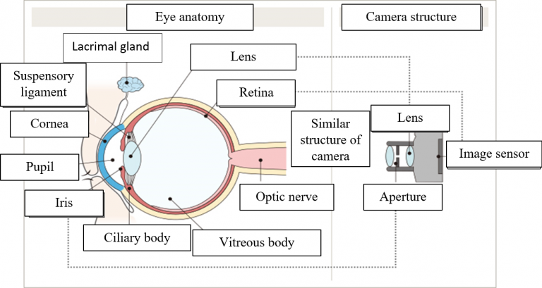
The eyes are considered the most beautiful and modern camera in the world. The eye captures images similar to the way the camera does.
To capture an image, the light reflected from the object is refracted through the lens system and focused on the film. The light signal here causes chemical reactions on the film, and then produces pictures through photo making process.
Likewise, the eye has a “lens” system consisting of cornea and lens. The light will, after being refracted through the cornea and lens, focus on the retina. The light signal here will be converted into neural signals by photoreceptor cells on the retina. That signal is then transmitted to the brain via the optic nerves and identified as imaging in the brain. This is how the eye works to see something.
For the camera, we have to adjust to obtain precise focus and light level for sharpness, and clean the dirty lens and maintain it carefully. In fact, our eyes perform those tasks completely automatically.
For example, to change focus, our lens will change its curvature controlled by the ciliary muscles. Adjusting the iris’ elasticity shall result in changes of the pupil’s size, which in turn controls the intensity of the incoming light beam. The main and auxiliary lacrimal glands work to keep the cornea lubricated, this is a natural cleaning and protection mechanism that is bestowed on the eyes by nature.
These operations take place automatically under the extremely sophisticated control of neural mechanisms, which no high-end camera can match.