.jpg)
Scleral buckling is a surgical procedure to repair a retinal detachment by removing the detached humor under the retina, then placing a small band or buckle around the outside of the eyeball to push the sclera toward the retinal tear and keep the retina in place.
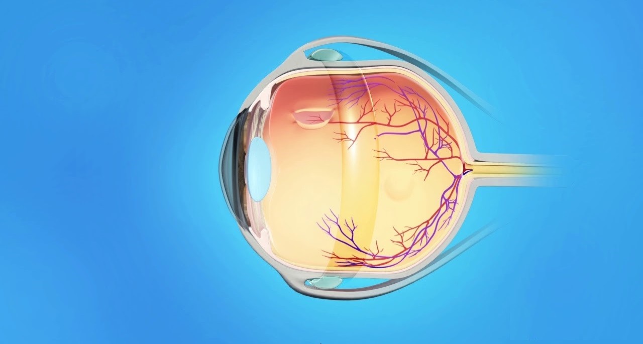
Retinal detachment is an eye condition in which the retina is detached from the tissue underneath. Its initial cause may be a small retinal tear/hole, letting the fluids flow under the retina, gradually causing the retina to be partly separated from the eye wall. If not treated quickly, the entire retina can be completely detached, leading to a permanent loss of vision. Currently, beside the surgical procedure, there is no effective treatment for retinal detachment.
Around 1 hour before the surgery, you shall be provided with antibiotics, anti-inflammatory and pupil dilation drops every 3-5 minutes.
Step 1: Local anesthetic injection near the eyeball
The eye is cleaned with an antiseptic solution; then local anesthetic drops are given first to numb the surface of the eye and anesthetic injection is given near to the eye, but not the eyeball itself. You may first feel stinging or pressure which usually lasts within 3-5 minutes.
Step 2: Perform scleral buckling
– Clean the eye(s) with an antiseptic solution.
– Cover it with sterile surgical drape.
– Place an eyelid holder.
– Separate conjunctiva to reveal the orthogonal muscles, insert thread to hold the orthogonal muscles, insert silicon buckles under the orthogonal muscles.
– Drain the sub-retina fluid through the sclera, then freeze the area by Cryo therapy.
– Sew to fix the silicone buckle on the sclera with surgical thread, insert expanding gases.
– Stitch the conjunctiva.
– Apply antibiotic eye drops.
– Apply bandage and complete the surgery.
In the scleral buckling, depending on your conditions, your doctor may also perform one of the following:
Cryopexy
In the retinal cryopexy, the surgeon will use a special probe to apply intense cold energy to freeze the area around the retinal tear, then this creates swelling that becomes scar tissue sealing the retina to the wall of the eye and preventing further damage or a complete retinal detachment caused by the overflow of the humor under the retina. This method is as effective as the intraocular laser.
Insert expanding gases
After the vitreous humor removal and retinal procedures are performed, the doctor will inject fluid (BSS solution) into the vitreous body and complete the surgery. There are retinal diseases that require the retinal tamponade, at that time the doctor can inject gases to hold the retina in position. In this case, patient will be limited in resting position and find it difficult to see for about 10-14 days after surgery. It is highly important to follow the post-operative instructions.
For those with retinal detachment, if their surgery has not been performed immediately, they should limit activities such as taking a flight or do vigorous activities. Besides basic needs, they should lie down in bed and avoid too many activities. Other notices:
- Having breakfast normally on the day of surgery (except for those with diabetes).
- Should not use alcohol or stimulants before the surgery.
- Continuing to use daily prescription drugs if any.
- Should not make up and use cosmetics, keep the eyes and face clean on the day of surgery.
- Should not wear tight pullovers or shirts made of fur.
- Should not wear items such as jewelry, watches or hairpins into the operating room.
- Getting enough sleep and stay comfortable before the day of surgery.
- Should not drive by themselves on the day of surgery.
After the surgery, you are asked to maintain a face-down position for 3-5 days. You can stand up, and walk normally for basic needs such as eating and hygiene.
Post- cryopexy inflammation takes at least 10-14 days to heal. During this time, the risk of retinal detachment is very high, so you are recommended to take full rest. In addition, you will have been applied with pupil dilation drops to facilitate examination during your hospitalization (at least 3 days), so you will not see clearly during this time.
The scleral buckling is aimed at preventing serious development of the retinal tear or detachment, in order to maintain vision as it was before the surgery for as long as possible. Due to the complexity of natural inclination of each person and the disease severity, the surgery outcome and postoperative vision cannot be fully prognosed. Therefore, it is not possible to guarantee the postoperative results with certainty. The post-operative vision can be improved, but very little, or cannot. The eye with retinal detachment, even if treated by surgery, cannot return to its original (pre-retinal detachment) condition.
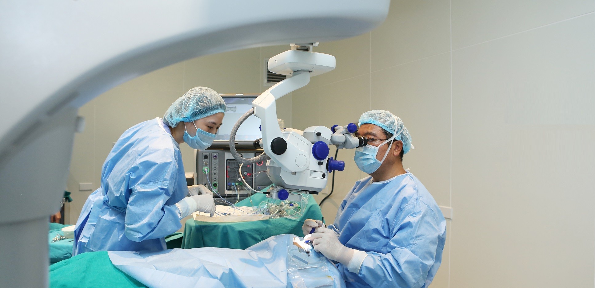
Experienced surgeons
The scleral buckling at our Japan International Eye Hospital are carried out by the surgeons with many years of experience in retina & vitreous diseases.
Quick, strict and professional procedure
Our rigorous and thorough pre-operative intensive examination process, which meets the Japanese standards, enables us to have accurate diagnosis and treatment indication in time. We deliver a comprehensive care service, supporting you throughout your surgery. You can visit us for an examination or surgery alone. Convenient procedure, minimizing waiting time in all examination steps.
*Note: The treatment performances may vary depending on the physical conditions/ natural disposition of each person. You should get examination and advice as well as direct instructions by your doctor before making a decision for any surgery or procedure.