Retinopathy
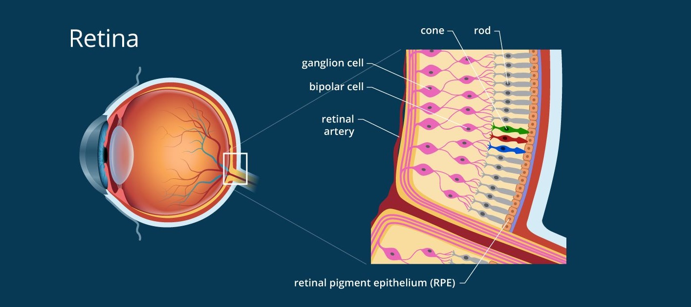
1. What Is Retina?
2.1 Retinal tear
Retinal tear is the main cause of retinal detachment. It is usually caused by aging of the eye. The eye contains a transparent tissue called vitreous humor which contacts the retina and lens surface, but there will be a gap gradually formed on this interface. It is because the vitreous humor is aged and emulsified to be more liquid, resulting in its gradually decreased volume. When this occurs, the vitreous humor and retina separate from each other, causing a posterior vitreous detachment. The posterior vitreous detachment is a condition that can happen to anyone and can cause retinal traction and then retinal tear.
Subjects at risk of retinal tear:
- Retinal tear may occur due to natural aging of the eye
- People with severe myopia are also at high risk of retinal detachment.
- Retinal tears can also be caused by physical effects, such as bumps in the eyes while playing sports.
Symptoms of retinal tear:
- Sudden onset of black spots or ‘floaters’ in your field of vision
- Photopsia (flashes of light) in one or both eyes
- A curtain-like shadow over your visual field
- Distorted vision
In the place of retinal tears, water can flow into the back of the retina through this fistula, causing retinal detachment. If not treated promptly, this condition may cause serious vision impairment; hence, examination and treatment of retinal tear is extremely essential.
How to treat retinal tear
Upon detection of retinal tear, the Laser photocoagulation procedure may be applied for photocoagulation of the torn retina area, preventing its progression. But, even with Laser photocoagulation, it still takes a few weeks to seal the retina, therefore, patients are recommended to follow the doctor’s instructions to achieve the best result.
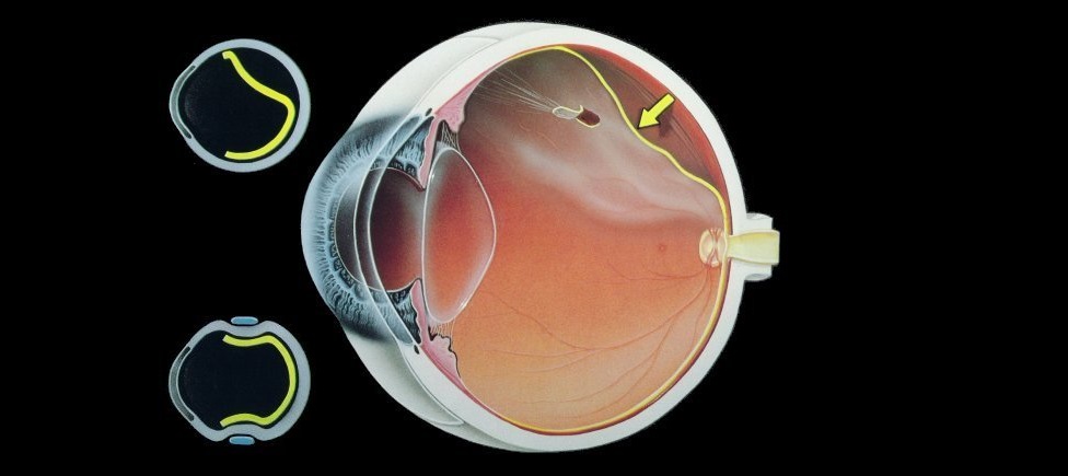
2.2 Retinal detachment
Retinal detachment occurs when the retina has tear(s). The vitreous humor then may pass through the retinal tear and enter beneath the retina, gradually lift the retina off its beneath tissue, disrupting the nutrient supply process.

Symptoms of Retinal detachment
- Blurred vision
- Sudden onset of black spots or ‘floaters’ in your field of vision
- Distorted vision
Subjects at high risk of retinal detachment
- Elderly, aging
- Severe myopia
- Complications of diabetes, kidney disease, preeclampsia
How to treat retinal detachment
The treatment of retinal detachment depends on its situation. It may include vitrectomy and scleral buckling.
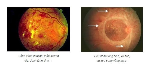
2.3 Diabetic retinopathy
One of the complications of diabetes is diabetic retinopathy. This is one of the leading causes of blindness in the world.
When blood sugar levels rise, blood viscosity increases, increasing pressure on the vessels. Because the retina is a concentrated place of various capillaries, blockage or breaking of these blood vessels may cause retinal hemorrhage.
Thediabetic retinopathy can be divided into 3 stages as follows:
- Retinopathy of prematurity (early stage)
- Pre-proliferative retinopathy (second stage)
- Proliferative retinopathy (third stage)
Symptoms of the diabetic retinopathy:
- In the early stages, the patient hardly has any obvious symptoms
- In stage three, symptoms included: impaired vision, the patients may see “black floaters” in their field of vision
How to treat diabetic retinopathy
The treatment of the diabetic retinopathy depends on its situation. Doctors can prescribe medication, laser, or surgery.
2.4 Retinal vein occlusion (RVO)
This is a common condition in the elderly, related to blood pressure and atherosclerosis. In the retina, arteries run across veins, membranes of the blood vessels are in contact. If patients have atherosclerosis caused by high blood pressure, the arteries compress the veins, causing blood blockage in the veins and blood overflow. This overflow causes retinal hemorrhage or accumulates in the retina to form edema points.
Retinal vein occlusion can be divided into two categories, namely:
- Branch Retinal Vein Occlusion (BRVO)
- Central Retinal Vein Occlusion (CRVO)
Symptoms of Retinal vein occlusion
- Retinal venous occlusion often has sudden onset in one eye. Its symptoms range from gentle to very severe depending on the location and severity of the vein occlusion. The patient may even have no symptoms at first, or feel like there is a dark spot in one part of the eye, without affecting vision.
- But usually, the patient suddenly has poor vision, feeling like looking through the fog, or as if there are black patches in front of his eyes, which can lead to severe vision impairment within minutes or 2-3 days.
Subjects at risk of Retinal vein occlusion
- Those with high blood pressure, diabetes, cardiovascular disease, dyslipidemia or eye diseases such as glaucoma, vasculitis.
2.5 Pigmentary retinal degeneration
The retina is made up of about 100 million photoreceptor cells called “photoreceptors”. When those photoreceptors cease to function, they cannot sense light and send images to the brain. Pigmentary retinal degeneration is a condition that occurs in both eyes when those photoreceptors degenerate and lose their function.
Symptoms of the Pigmentary retinal degeneration
- Impaired vision at night or in a low light environment.
- Narrow visual field or peripheral vision loss: The patient will feel like looking in a tunnel (called tubular vision) and often slip.
- Peripheral vision loss – In severe cases, loss of both central and peripheral visual field can happen.
- Its development is not the same in all patients. The symptom also depends on its stage.
Causes of the Pigmentary retinal degeneration
Causes leading to the pigmentary retinal degeneration are currently unknown. However, some experts believe that almost retinal pigment degeneration is related to genetic abnormalities, and think that its severity and progression rate depend on the level of genetic mutation, how it is inherited.
The genetic mutation can be inherited from a parent, or both parents, in which the recessive gene accounts for about 60-70%, the dominant gene is about 25%, the rest is on the X chromosome gene.
A child with a parent having the pigmentary retinal degeneration may be at higher risk of suffering this disease.
How to treat the Pigmentary retinal degeneration
Currently, there is no treatment has been proven the effectiveness in treating the pigmentary retinal degeneration. Some methods that can slow its development include adding an appropriate vitamin A as prescribed by your doctor, using supportive devices such as magnifying glasses, night vision binoculars, daily sunglasses to protect the retina and taking regular exams for the accurate assessment.
By examination, in case of detecting any retinal abnormalities, the doctor will recommend treatment based on its conditions as follows:
Intravitreal Anti-VEGF Injection
For retinal damages in their early stages, the patient will be treated by intravitreal injection of anti-vascular endothelial growth factor inhibitor to maintain and improve vision, and prevent the retinal detachment.
Laser photocoagulation
In the event that retinal pathologies make the retina too thin and have the risk of tearing at any time, the patient shall undergo a laser photocoagulation to seal the retina firmly, limit the retinal tear and detachment.
In cases that the retina has tears, small detachments, the patient should get the laser photocoagulation on the same day of examination to seal the tear, prolonged un-treatment may result in total retinal detachment and complete loss of vision.
Vitrectomy
In a more severe stage, when retinal damage cannot be improved by the intravitreal injection and laser photocoagulation, the doctor will use vitrectomy to treat retinal detachment or retinal hemorrhage to keep vision of the patients.
Vitreous humor is a transparent mass of gel behind the lens that fills 80% of the eyeball space. It acts to transmit the light rays from outside to the back of the eye, providing nutrients for the lens and retina. It also creates pressure to fix the lens shape, helping the lens to focus a sharp image on the retina. Vitrectomy is a surgery to remove part of or entire vitreous humor for the purpose of treating the retinal diseases.
The retinal diseases may appear without any symptoms or with vague symptoms, but they have very quick development and may lead to complete loss of vision. To detect retinal abnormalities at an early stage, regular eye exams or general health checks are not sufficient. Therefore, patients need to actively carry out screening of those diseases for early detection and treatment.