DISEASES THAT LEAD TO BLINDNESS - RECOGNITION, PREVENTION & TREATMENT | JAPAN INTERNATIONAL EYE HOSPITAL
Eyes are the important and essential part of every person, that needed to be cared for and worthy of investment. Protect your eye health today with Japan International Eye Hospital by recognizing, preventing and treating diseases that cause blindness.

Cataract
Cataracts (also known as dry cataracts, cataracts) is a phenomenon of protein concentration in clusters, creating opaque areas in the lens, scattering light passing through, blocking light from reaching Retinal. Cataracts make the patient's vision impaired, blurred vision and if not treated promptly, can lead to blindness. In addition, mature cataracts can cause glaucoma, red eyes, sore eyes, and headaches. At that time, not only is the patient permanently blind, but the eyeball may also need to be removed.

Symptoms of Cataracts
Signs and symptoms of cataracts include:
- The phenomenon of blurred vision: The patient will feel "it seems" that everything is slightly blurred, like there is a thin film of fog in front of the eyes.
- Reduced night vision: As cataracts progress, the lens becomes darker, affecting vision in low light, especially at night.
- Glare: Patients often feel glare when looking at light sources, glare, even pain.
- Reduced color perception: The color of objects that the eye perceives becomes darker, grayer than reality.
- Halos appear everywhere: Cataracts can cause light diffraction, so people with light bulbs often see halos around them.
- Progressive myopia beyond the age of 40: Continuous lens replacement with an increasing number of degrees can be one of the manifestations of cataracts.
- Double vision, triple vision: Diffraction from cataracts can cause double or triple vision.

In the initial stage, only a small part of the lens is cloudy, has not significantly affected vision, patients often cannot recognize abnormalities on the eyes. As the disease progresses, the cloudy portion of the lens becomes wider, resulting in distortion of light passing through the lens. By this time, the new signs are more obvious, and the person begins to notice and realize they have the disease.
How to prevent cataracts
- Eat a healthy diet: Some studies show that foods rich in antioxidants like vitamins C and E, and Lutein and Zeaxanthin can help prevent cataracts.
- Limit tobacco use: Smoking reduces the oxygen concentration in the lens, which in turn causes the lens to gradually lose its natural transparency and turn opaque. The highly toxic heavy metal cadmium in tobacco tends to accumulate in the vitreous nucleus, contributing to accelerated disease formation.
- Limit alcohol use: Drinking alcohol causes the metabolism and excretion to become overloaded. High blood alcohol levels for a long time continuously will cause poisoning and affect many organs in the body including the eyes.
- Wear sunglasses: Studies show that ultraviolet light from the sun is a major contributor to the development of cataracts. The American Optometric Association recommends sunglasses with the following criteria: labeled to block 100% of UV rays, block both UV A and B rays, and have wide-rimmed frames to prevent light from entering your eyes from behind. beside.

- Checking blood sugar with patients with diabetes: Unstable blood sugar over time will cause blood vessels to narrow, thrombosis occurs, and the blood transport to the capillaries of the eye area is slow. . Long-term damage to blood vessels will cause cataracts.
Cataract treatment methods
- Phaco surgery: It is the process of pulverizing the cloudy lens with high-frequency ultrasound and sucking it out. It is then replaced with a suitable artificial lens to restore vision.
- Cataract Laser Surgery: Has the same mechanism of pulverizing the cloudy lens to take it out and replace it with an artificial lens suitable for the patient's eyes like Phaco method. The difference is that the Femtosecond Laser is put into use and replaces a number of manual operations such as making incisions, creating anterior capsular tear, and assisting in splitting the cloudy lens.

👉 Learn more about Cataract treatments here.
Glaucoma
Glaucoma is a term used to describe a group of diseases that are characterized by nerve damage and loss of vision. The disease often appears silently, causing damage to the eyes, if not detected and treated promptly can lead to blindness.

Symptoms of Glaucoma
Signs of glaucoma will depend on the type of disease.
- With chronic open-angle glaucoma (COAG): The disease appears silently, progresses slowly, patients often do not feel any recognizable signs and only come to the doctor when the disease has progressed severely. . Some mild symptoms may include eye strain, vision like a thin fog in front of your eyes in the morning, transient blurred vision when working with your eyes a lot, when nervous, when you worry a lot.
- With acute angle-closure Glaucoma: Patients with glaucoma often appear in the evening, after working tired, after anxiety or have mental concussion. In addition, patients may also experience severe eye pain, red eyes, headache (on the same side as the affected eye), blurred vision, halos around lights, nausea.
- With congenital glaucoma in children: Patients will experience symptoms of watery eyes, sensitivity to light, eyelid twitching, increased corneal size (feeling that the eyes are heavier and more swollen). In addition, parents can also notice children's actions such as rubbing their eyes, squinting, and closing their eyes.
- With secondary glaucoma and other forms: The dependent symptoms of the disease will depend on the cause of the glaucoma in the eye. For people with uveitis, symptoms may include seeing halos and being afraid of bright light. For people with trauma to the eye such as corneal edema, bleeding or retinal detachment, the symptoms of glaucoma may be more difficult to detect. If the cause of the glaucoma is a cataract, the patient's vision may be impaired for a long time. If you have a history of eye trauma, severe cataracts, or inflammation in your eye, your doctor will check to make sure your eyes don't have glaucoma. Another common cause of secondary glaucoma is the use of topical or systemic steroids.
How to prevent Glaucoma
The American Optometric Association (AOA) has listed some good habits that can reduce the risk of developing and progressing glaucoma:
- Exercise regularly.
- Eat more vegetables.
- Avoid upside down positions.
- Protect your eyes from the sun.
- Monitor and control BMI.
- Cut down or even eliminate caffeine.
- Quit smoking.

Glaucoma treatments
- Use eye drops: Helps lower eye pressure by increasing the drainage of aqueous humor out of the eye or reducing the amount of eye fluid secreted.
- Use oral medication: Helps reduce secretions (water) in the eye. Some commonly used drugs: beta-blocker, carbonic anhydrase inhibitor.
In case the treatment of Glaucoma with the use of drugs does not achieve the desired effect, the doctor will appoint the patient to perform surgery or laser treatment. The purpose of these methods is to create an outlet for the aqueous humor, stabilizing the pressure in the eye. Depending on the condition as well as the disease, there will be different laser methods including:
- Laser cutting of the peripheral iris.
- Laser shaping the raft.
- Laser photocoagulation of the ciliary body.
- Corneal trabeculectomy surgery.
- Non-perforating deep sclerectomy.
- Surgical placement of anterior chamber drain valve.
👉 Learn more about Glaucoma treatments here.
Retinal detachment
The retina is considered one of the most important parts of the eye. Even a very small damage to the retina can have significant effects on vision.
Retinal detachment is a condition in which the innermost and rear membranes of the eye separate from the remaining layers, causing partial loss of vision or permanent blindness if not treated promptly. The progression from detection of retinal tears or partial retinal detachment to total retinal detachment can be very rapid in just a few weeks or even days.

However, most of the time it is difficult for patients to recognize the symptoms of retinal detachment and only when their vision is severely impaired will they visit an eye specialist. Depending on the type of disease, the degree of disease that affects vision will also be different.
Signs of retinal detachment
Signs of retinal detachment depend on the stage of the disease.
- Before retinal detachment occurs: Signs are not clear, peripheral vision gradually decreases over several days or weeks, flies in front of the eyes occur more often, seeing flashes of light and blurred vision.
- When retinal detachment occurs: Part of the vision is gray or dark as if there is a veil in front of the eye.
How to prevent retinal detachment
- Avoid rubbing your eyes.
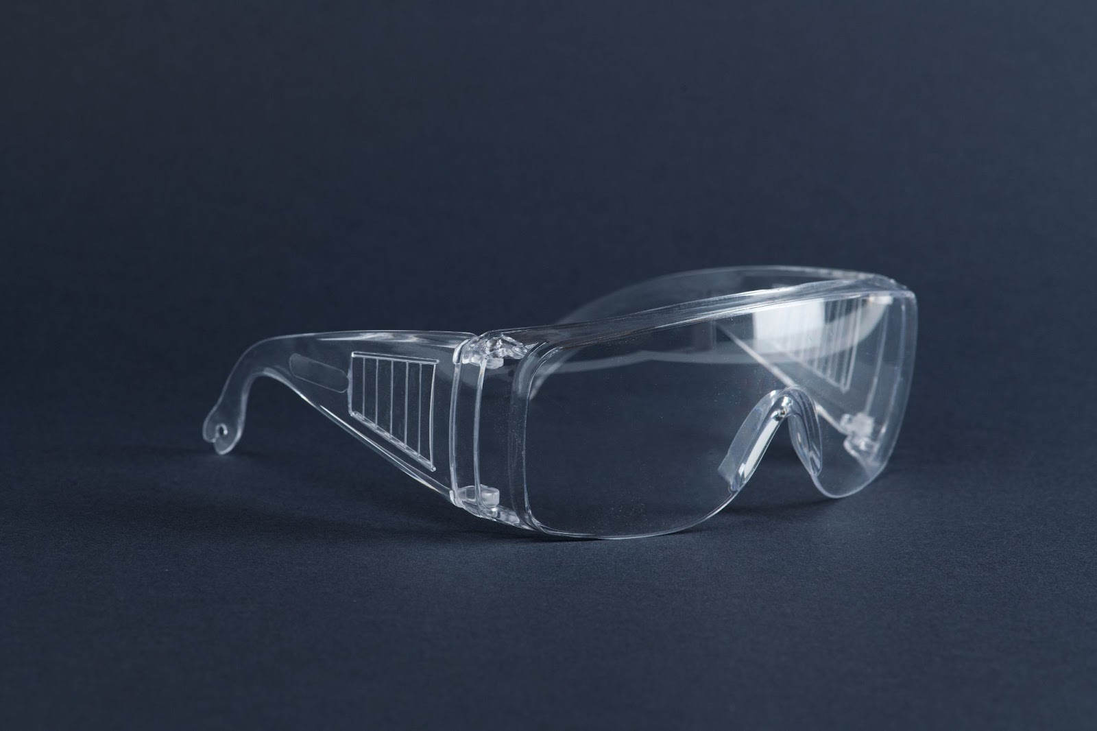
- Minimize the risk of eye injury by wearing protective eyewear.
- Subjects in the high-risk group for retinal detachment such as severe myopia, retinal degeneration, diabetic retinopathy, etc., should have periodic visits at least every 6 months to screen for risks and treat timely if needed.
- Visit your doctor right away if you notice any unusual symptoms such as:
- flies fly a lot, see flashes of light, partial vision looks like gray or black, blurred vision.
Treatment methods for retinal detachment
- Vitrectomy: A method to remove part or all of the vitreous in the eye to serve the treatment of pathologies on the retina. This method is indicated for the treatment of pathologies such as vitreous hemorrhage, proliferative retinopathy, complicated retinal detachment, endophthalmitis, ...
- Belt implant surgery: A method of treating retinal detachment with the mechanism of removing the detachment, causing inflammation of the retinal tear, pressing the convex sclera towards the vitreous chamber to seal and cause an inflammatory reaction to scar the tear. The retina causes the retina to press flat against the wall of the eyeball. The method is indicated for patients with retinal detachment with a tear in the peripheral retina.
- Laser retinal photocoagulation: Laser retinal photocoagulation is a procedure that uses a laser with a wavelength suitable for the absorption spectrum of the retinal pigment epithelium to coagulate the cell layers, causing sticky scars between the melanocytes. retina and retina. The method is indicated for retinal degeneration due to complications of diabetes or photocoagulation of tears on the retina, limiting the risk of retinal detachment, fundus bleeding.

👉 Learn more about the treatments for Retinopathy here.
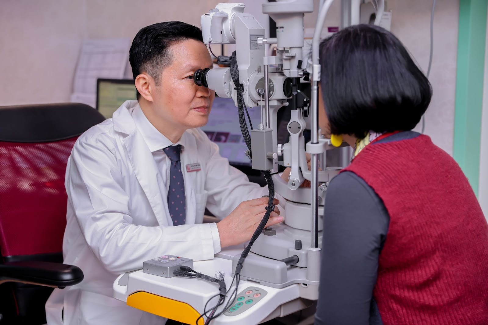
To prevent diseases that cause blindness, or any other eye disease, proactively have regular eye exams every 6 months to monitor eye health and take timely intervention measures.
👉 Register for an eye exam at Japan International Eye Hospital: https://jieh.vn/en/make-an-appointment